Evidence-Based Management of Upper Extremity Osteoarthritis: From Evaluation to Arthroplasty Rehab
Gain practical strategies to evaluate and manage upper extremity osteoarthritis. This article covers conservative care, surgical decision-making, and rehabilitation after arthroplasty.
October 2, 2025
11 min. read
Osteoarthritis (OA) of the upper extremity may not be as common as hip or knee OA, but its impact on function, independence, and quality of life can be just as profound. For many older adults, progressive pain and stiffness in the shoulder, elbow, wrist, or hand translates into difficulty with overhead reaching, grooming, lifting, or even simple daily tasks.
In this article, we’ll focus on two central themes in upper extremity OA: evaluating and managing glenohumeral osteoarthritis, and rehabilitation following shoulder arthroplasty. We’ll also briefly touch on the elbow, wrist, and hand, and share a clinical case example.
Clinical evaluation of shoulder osteoarthritis
Shoulder osteoarthritis, particularly glenohumeral osteoarthritis, is a condition most often seen in adults over 65. Patients typically report a gradual onset of pain and stiffness that worsens with overhead activities or after rest. Many also describe morning stiffness, difficulty reaching into cabinets, or pain when lying on the affected side at night.
Prevalence data underscore the importance of screening: radiographic changes appear in 15 to 20 percent of adults, and women are more frequently affected than men. Occupations and sports with repetitive overhead demands further elevate risk.1
Key clinical features include:
Progressive upper arm and shoulder pain (distinct from neck pain)
Morning stiffness or stiffness after rest
Decreased range of motion in all planes, especially internal rotation
Crepitus or joint noises with movement
Weakness, often linked to rotator cuff involvement
When evaluating a patient, it is essential to distinguish OA from other conditions with overlapping presentations. Common differential diagnoses include:
Adhesive capsulitis, which develops over weeks rather than years
Rotator cuff tears, often associated with trauma in younger patients or attritional wear in older adults
Subacromial pain syndrome, more common in younger, active populations
Glenohumeral instability, typically following a history of dislocation or subluxation
Imaging serves as a confirmatory tool rather than the primary basis for diagnosis. The American College of Radiology recommends plain radiographs as the most appropriate first-line study. MRI can be helpful to confirm the diagnosis and rule in pathology, but it is less effective for ruling it out.
By combining careful history-taking, focused examination, and judicious use of imaging, you can confidently identify glenohumeral OA and tailor appropriate management strategies.
Non-surgical management strategies
Not every patient with shoulder osteoarthritis is ready—or appropriate—for surgery. Many are looking for practical, nonoperative strategies to stay active and delay more invasive interventions.
The APTA Clinical Practice Guideline for Glenohumeral OA emphasizes that exercise-based rehabilitation should remain the foundation of care, supported by patient education and adjunctive options when appropriate. International recommendations, such as the OARSI guidelines, echo this approach, underscoring the value of a multimodal, individualized plan.
Here are several strategies supported by guidelines and clinical consensus:2,3
Lifestyle modification
Adjusting activities to reduce overhead strain, heavy lifting, or repetitive motions that provoke symptoms. Even small changes in how patients approach daily tasks can ease joint stress and prolong function.
Physical therapy
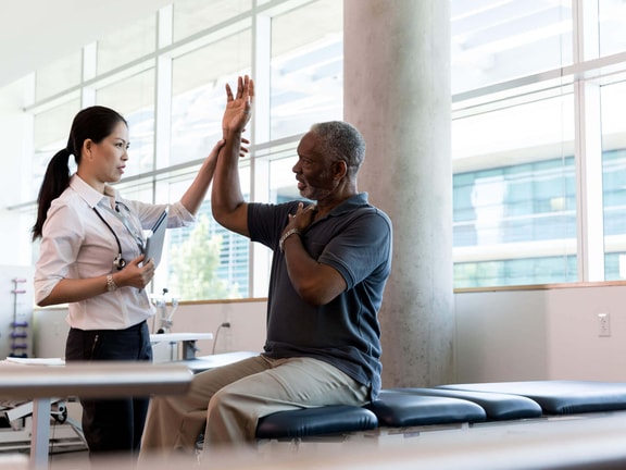
A program built on exercise forms the basis of non-surgical management. This may include modalities for pain relief, joint mobilization, range-of-motion activities, and progressive strengthening. Programs should be tailored to the individual’s tolerance and functional goals.
Injections
Corticosteroid or hyaluronic acid injections can provide short-term symptom relief, especially for patients experiencing acute flare-ups. They are not curative but may improve tolerance for activity and rehabilitation.
Medications
Nonsteroidal anti-inflammatory drugs (NSAIDs), aspirin, or acetaminophen may be used to manage pain and help patients better participate in therapy. These should be considered supportive measures alongside an active rehab program, rather than standalone treatments.
Non-surgical care will not reverse the degenerative process, but it can reduce pain, improve mobility, and meaningfully delay surgical intervention. The therapist’s role is to monitor outcomes, adapt the plan as needed, and help patients maintain engagement in meaningful daily activities for as long as possible.
The rise of shoulder arthroplasty
For some patients with severe shoulder osteoarthritis, non-surgical care simply isn’t enough. When pain interferes with sleep, daily activities, and quality of life despite months of therapy and medication, surgery becomes an important next step.
Over the last two decades, shoulder arthroplasty has increased substantially, with reverse total shoulder arthroplasty (rTSA) accounting for much of the growth due to its effectiveness in cuff tear arthropathy.4
Indications for shoulder arthroplasty include:
Degenerative joint disease (osteoarthritis, rheumatoid arthritis, or post-traumatic arthritis)
Proximal humeral fractures
Massive rotator cuff tears leading to cuff tear arthropathy
Avascular necrosis
Failed previous replacements
Within these cases, surgeons decide between two main procedures:
Anatomic total shoulder arthroplasty (ATSA): Restores normal biomechanics and requires an intact rotator cuff.
Reverse total shoulder arthroplasty (rTSA): Reverses the ball-and-socket orientation, allowing the deltoid to take over when the rotator cuff is deficient.
Both procedures share the same goals: reducing pain, restoring function, and improving sleep quality. For many patients, better rest is one of the most meaningful outcomes.
Rehabilitation after shoulder arthroplasty
Surgery is only the first step in restoring function. For patients who undergo shoulder arthroplasty, outcomes depend heavily on rehabilitation.
General principles apply across both procedures. Patients are typically immobilized in a sling for four to six weeks, with restrictions on movements that could jeopardize repair, such as excessive external rotation or combined extension with internal rotation. Early priorities include pain management, joint protection, and gentle mobility. Therapy then progresses in phases, gradually layering motion, strengthening, and functional activity as healing permits.
For anatomic total shoulder arthroplasty (ATSA):5,6
Phase 1 (0–4 weeks): Pain and inflammation control, passive range of motion, scapular isometrics.
Phase 2 (4–8 weeks): Assisted and active range of motion, gentle isometrics, scapular strengthening.
Phase 3 (8–12 weeks): Resisted strengthening, manual therapy, light functional activities.
Phase 4 (12+ weeks): Full active range of motion, resistive training, and gradual return to advanced activities.
For reverse total shoulder arthroplasty (rTSA):7
Early progression is more conservative, with passive range of motion often delayed until weeks 4–6 to allow deltoid and soft tissue healing.
Rehabilitation emphasizes deltoid and scapular stabilizer strength, since these muscles compensate for the deficient rotator cuff.
Patients may return to light household activities around 12 weeks, but many surgeons recommend lifelong lifting restrictions to protect the prosthesis.
The evidence base for rehabilitation after ATSA is supported by systematic reviews and consensus statements, while guidelines for rTSA are more variable and often based on expert opinion. Despite these gaps, phased progression—combined with patient education and careful monitoring—remains the most widely accepted approach.
Case example: Glenohumeral OA to reverse TSA
A 68-year-old woman presents with a long history of progressive shoulder pain and stiffness. She reports morning stiffness, crepitus with motion, and increasing difficulty reaching overhead. On examination, her range of motion is limited in all planes—particularly internal rotation—and there is weakness consistent with rotator cuff involvement.
Her care begins with conservative management, including activity modification, joint mobilization, and progressive strengthening. These interventions provide short-term relief, but her symptoms return over time. Imaging later confirms advanced joint degeneration with significant rotator cuff pathology.
Given the extent of her condition, she undergoes a reverse total shoulder arthroplasty. Early rehabilitation emphasizes sling protection and a cautious introduction of passive range of motion at week four. By week eight, she progresses to assisted and active motion with scapular stabilization. At four months, she achieves functional overhead elevation, resumes light activities such as gardening, and reports a marked reduction in pain.
This example illustrates the full continuum of care: starting with conservative approaches to managing symptoms, and transitioning to surgery and postoperative rehabilitation when function and quality of life are no longer sustainable without them.
Beyond the shoulder: Elbow, wrist, and hand OA
Although less common than shoulder osteoarthritis, elbow, wrist, and hand OA can significantly affect function and independence. Each joint has unique risk factors and management strategies, but individualized, evidence-informed care principles apply across all three.
Elbow OA
More common in men, often linked to manual labor or overhead sports.
Conservative care mirrors that of the shoulder: activity modification, joint mobilization, and strengthening.
Advanced cases may require arthroscopic debridement or, in severe cases, arthroplasty.8
Wrist OA
Often presents with pain and instability that interferes with daily tasks or weightbearing through the hand.
First-line care includes splinting, joint mobilization, and therapeutic exercise.
Surgery is generally reserved for persistent instability or functional loss.
Hand OA
Particularly common at the carpometacarpal (CMC) joint.
The European League Against Rheumatism (EULAR) recommend exercise, orthoses, and topical NSAIDs as effective nonoperative strategies.9
Across these joints, clinicians should balance pain relief, preservation of function, and patient goals.
Next steps in evidence-based OA care
Upper extremity osteoarthritis challenges clinicians to think beyond the hip and knee. Early recognition of glenohumeral OA, thoughtful application of non-surgical strategies, and structured rehabilitation after arthroplasty are all critical in helping patients regain motion, reduce pain, and improve quality of life. The evidence base continues to evolve, but current clinical practice guidelines, expert consensus, and case-by-case decision-making provide a strong framework for practice.
If you’re looking to broaden your expertise, there’s much more to explore. My Medbridge courses provide deeper insight into osteoarthritis across multiple joints—covering differential diagnosis, surgical decision-making, and evidence-based rehabilitation strategies:
References
Kobayashi, T., Takagishi, K., Shitara, H., Ichinose, T., Shimoyama, D., Yamamoto, A., Osawa, T., & Tajika, T. (2014). Prevalence of and risk factors for shoulder osteoarthritis in Japanese middle-aged and elderly populations. Journal of shoulder and elbow surgery, 23(5), 613–619. https://pubmed.ncbi.nlm.nih.gov/24561177/
Michener, L. A., Heitzman, J., Abbruzzese, L. D., Bondoc, S. L., Bowne, K., Henning, P. T., Kosakowski, H., Leggin, B. G., Lucado, A. M., & Seitz, A. L. (2023). Physical Therapist Management of Glenohumeral Joint Osteoarthritis: A Clinical Practice Guideline from the American Physical Therapy Association. Physical therapy, 103(6), pzad041. https://pubmed.ncbi.nlm.nih.gov/37115808/
Bannuru, R. R., Osani, M. C., Vaysbrot, E. E., Arden, N. K., Bennell, K., Bierma-Zeinstra, S. M. A., Kraus, V. B., Lohmander, L. S., Abbott, J. H., Bhandari, M., Blanco, F. J., Espinosa, R., Haugen, I. K., Lin, J., Mandl, L. A., Moilanen, E., Nakamura, N., Snyder-Mackler, L., Trojian, T., Underwood, M., … McAlindon, T. E. (2019). OARSI guidelines for the non-surgical management of knee, hip, and polyarticular osteoarthritis. Osteoarthritis and cartilage, 27(11), 1578–1589. https://pubmed.ncbi.nlm.nih.gov/31278997/
Dillon, M. T., Inacio, M. C., Burke, M. F., Navarro, R. A., & Yian, E. H. (2013). Shoulder arthroplasty in patients 59 years of age and younger. Journal of shoulder and elbow surgery, 22(10), 1338–1344. https://pubmed.ncbi.nlm.nih.gov/23571084/
Thigpen, C. A., Shaffer, M. A., Gaunt, B. W., Leggin, B. G., Williams, G. R., & Wilcox, R. B., 3rd (2016). The American Society of Shoulder and Elbow Therapists' consensus statement on rehabilitation following arthroscopic rotator cuff repair. Journal of shoulder and elbow surgery, 25(4), 521–535. https://pubmed.ncbi.nlm.nih.gov/26995456/
Wilcox, R. B., Arslanian, L. E., & Millett, P. (2005). Rehabilitation following total shoulder arthroplasty. The Journal of orthopaedic and sports physical therapy, 35(12), 821–836. https://pubmed.ncbi.nlm.nih.gov/16848103/
Boudreau, S., Boudreau, E. D., Higgins, L. D., & Wilcox, R. B., 3rd (2007). Rehabilitation following reverse total shoulder arthroplasty. The Journal of orthopaedic and sports physical therapy, 37(12), 734–743. https://pubmed.ncbi.nlm.nih.gov/18560182/
Del Core, M. A., & Koehler, D. (2023). Elbow arthritis. Journal of Hand Surgery (American Volume), 48(6), 603–611. https://www.jhandsurg.org/article/S0363-5023(23)00003-5/abstract
Kloppenburg, M., Kroon, F. P., Blanco, F. J., Doherty, M., Dziedzic, K. S., Greibrokk, E., Haugen, I. K., Herrero-Beaumont, G., Jonsson, H., Kjeken, I., Maheu, E., Ramonda, R., Ritt, M. J., Smeets, W., Smolen, J. S., Stamm, T. A., Szekanecz, Z., Wittoek, R., & Carmona, L. (2019). 2018 update of the EULAR recommendations for the management of hand osteoarthritis. Annals of the rheumatic diseases, 78(1), 16–24. https://pubmed.ncbi.nlm.nih.gov/30154087/
Below, watch Christopher Bise discuss history and physical examination in glenohumeral osteoarthritis in this brief clip from his Medbridge course "Evidence-Based Management of Upper Extremity Osteoarthritis."

Meet the Author


Subscribe to Our Newsletter
Related Posts

November 18, 2024
Top Exercises for Shoulder Pain
By Medbridge

January 10, 2025
Top 5 Exercises for Hip Osteoarthritis
By Medbridge

January 31, 2025
Download a Free Shoulder Impingement Exercises PDF
By Medbridge

June 2, 2021
Shoulder Replacement: Addressing Shoulder Hike and Shrug
By John O'Halloran